Circulatory organs. Type Chordates. Superclass Pisces Meaning of the human circulatory system briefly
The circulatory system is a single anatomical and physiological formation, the main function of which is blood circulation, that is, the movement of blood in the body.
Thanks to blood circulation, gas exchange occurs in the lungs. During this process, carbon dioxide is removed from the blood, and oxygen from the inhaled air enriches it. Blood delivers oxygen and nutrients to all tissues, removing metabolic (decay) products from them.
The circulatory system is also involved in the processes of heat transfer, ensuring the vital activity of the body in different environmental conditions. Also, this system is involved in the humoral regulation of the activity of organs. Hormones are secreted by the endocrine glands and delivered to susceptible tissues. So the blood unites all parts of the body into a single whole.
Parts of the vascular system
The vascular system is heterogeneous in morphology (structure) and function. It can be divided into the following parts with a small degree of conventionality:
- aortoarterial chamber;
- vessels of resistance;
- exchange vessels;
- arteriovenular anastomoses;
- capacitive vessels.
The aortoarterial chamber is represented by the aorta and large arteries (common iliac, femoral, brachial, carotid, and others). Muscle cells are also present in the wall of these vessels, but elastic structures predominate, preventing their collapse during cardiac diastole. The vessels of the elastic type maintain the constancy of the blood flow velocity, regardless of pulse shocks.
Resistance vessels are small arteries, in the wall of which muscle elements predominate. They are able to quickly change their lumen, taking into account the needs of an organ or muscle for oxygen. These vessels are involved in maintaining blood pressure. They actively redistribute blood volumes between organs and tissues.
Exchange vessels are capillaries, the smallest branches of the circulatory system. Their wall is very thin, gases and other substances easily penetrate through it. Blood can flow from the smallest arteries (arterioles) into venules, bypassing capillaries, through arteriovenular anastomoses. These "connecting bridges" play a large role in heat transfer.
Capacitance vessels are so called because they are able to hold much more blood than arteries. These vessels include venules and veins. Through them, blood flows back to the central organ of the circulatory system - the heart.
Circles of blood circulation

Circulatory circles were described as early as the 17th century by William Harvey.
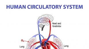
The aorta emerges from the left ventricle and begins the systemic circulation. The arteries that carry blood to all organs are separated from it. Arteries are divided into ever smaller branches, covering all the tissues of the body. Thousands of tiny arteries (arterioles) break up into a huge number of the smallest vessels - capillaries. Their walls are characterized by high permeability, so gas exchange occurs in the capillaries. Here, arterial blood is transformed into venous blood. Venous blood enters the veins, which gradually unite and eventually form the superior and inferior vena cava. The mouths of the latter open into the cavity of the right atrium.
In the pulmonary circulation, blood passes through the lungs. It gets there through the pulmonary artery and its branches. In the capillaries surrounding the alveoli, gas exchange with air occurs. Oxygenated blood flows through the pulmonary veins to the left side of the heart.
Some important organs (brain, liver, intestines) have peculiarities of blood supply - regional blood circulation.
The structure of the vascular system
The aorta, leaving the left ventricle, forms the ascending part, from which the coronary arteries are separated. Then it bends, and vessels depart from its arc, directing blood to the arms, head, and chest. Then the aorta goes down along the spine, where it divides into vessels that carry blood to the organs of the abdominal cavity, pelvis, and legs.
The veins accompany the arteries of the same name.
Separately, it is necessary to mention the portal vein. It carries blood away from the digestive organs. In addition to nutrients, it may contain toxins and other harmful agents. The portal vein delivers blood to the liver, where toxic substances are removed.
The structure of the vascular walls

Arteries have outer, middle and inner layers. The outer layer is connective tissue. In the middle layer there are elastic fibers that support the shape of the vessel, and muscle. Muscle fibers can contract and change the lumen of the artery. From the inside, the arteries are lined with endothelium, which ensures a smooth flow of blood without obstruction.
The walls of veins are much thinner than those of arteries. They have very little elastic tissue, so they stretch and fall off easily. The inner wall of the veins forms folds: venous valves. They prevent the downward movement of venous blood. The outflow of blood through the veins is also ensured by the movement of skeletal muscles, "squeezing out" the blood when walking or running.
Regulation of the circulatory system
The circulatory system almost instantly responds to changes in external conditions and the internal environment of the body. Under stress or stress, it responds with an increase in heart rate, an increase in blood pressure, an improvement in blood supply to the muscles, a decrease in the intensity of blood flow in the digestive organs, and so on. During rest or sleep, the reverse processes occur.
The regulation of the function of the vascular system is carried out by neurohumoral mechanisms. The highest level regulatory centers are located in the cerebral cortex and in the hypothalamus. From there, the signals go to the vasomotor center, which is responsible for vascular tone. Through the fibers of the sympathetic nervous system, impulses enter the walls of blood vessels.
In the regulation of the function of the circulatory system, the feedback mechanism is very important. In the walls of the heart and blood vessels there are a large number of nerve endings that perceive changes in pressure (baroreceptors) and the chemical composition of the blood (chemoreceptors). Signals from these receptors go to higher regulatory centers, helping the circulatory system quickly adapt to new conditions.
Humoral regulation is possible with the help of the endocrine system. Most human hormones in one way or another affect the activity of the heart and blood vessels. The humoral mechanism involves adrenaline, angiotensin, vasopressin and many other active substances. 
The subtype Cranial includes the only class of the Head Chordidae, which has only about 30-35 species of marine animals living in shallow water. A typical representative is Lancelet - Branchiostoma lanceolatum(genus Lancelet, class Headochord, subtype Cranial, type Chordata), the size of which reaches 8 cm. The body of the Lancelet is oval in shape, narrowed towards the tail, laterally compressed. Outwardly, the Lancelet resembles a small fish. Located on the back of the body tail fin in the form of a lancet - an ancient surgical instrument (hence the name Lancelet). Paired fins are absent. There is a small dorsal fin. On the sides of the body from the ventral side hang two metapleural folds, which fuse on the ventral side and form peribranchial, or an atrial cavity that communicates with the pharyngeal fissures and opens at the posterior end of the body with a hole - atriopore- outside. At the anterior end of the body near the mouth are the perioral tentacles, with which the Lancelet captures food. Lancelets live on sandy soils in the sea at a depth of 50-100 cm in temperate and warm waters. They feed on bottom sediments, marine ciliates and rhizopods, eggs and larvae of small marine crustaceans, diatoms, burrowing into the sand and exposing the front end of the body. More active at dusk, avoid bright lighting. Disturbed Lancelets swim quite quickly from place to place.
Covers. The body of the lancelet is covered skin, consisting of a single layer epidermis and thin layer dermis.
Musculoskeletal system. A chord stretches along the entire body. Chord- this is an elastic rod located on the dorsal side of the body and performing a supporting function. To the anterior and posterior ends of the body, the chord becomes thinner. The notochord protrudes into the anterior part of the body a little further than the neural tube, hence the name of the class - Cephalic. The notochord is surrounded by connective tissue, which simultaneously forms support elements for the dorsal fin and divides the muscle layers into segments using connective tissue
layers. Individual muscle segments are called myomers, and the partitions between them myoseptami. Muscles are formed by striated muscles.
body cavity at the lancelet secondary in other words, they are coelomic animals.
Digestive system. On the front of the body is oral hole, surrounded by tentacles(up to 20 pairs). The mouth opening leads to a large throat, which functions as a filtering apparatus. Through the cracks in the pharynx, water enters the atrial cavity, and food particles are sent to the bottom of the pharynx, where endostyle- a groove with a ciliary epithelium that drives food particles into the intestine. no stomach, but hepatic outgrowth, homologous to the liver of vertebrates. Medium intestine, without making loops, opens anal hole at the base of the tail fin. Digestion of food occurs in the intestines and in the hollow hepatic outgrowth, which is directed towards the head end of the body. Interestingly, the Lancelet retained intracellular digestion, intestinal cells capture food particles and digest them in their digestive vacuoles. This mode of digestion is not found in vertebrates.
Respiratory system. There are more than 100 pairs in the throat of the Lancelet gill cracks leading to peribranchial cavity. The walls of the gill slits are penetrated by a dense network of blood vessels in which gas exchange occurs. With the help of the ciliary epithelium of the pharynx, water is pumped through the gill slits into the peribranchial cavity and through the opening (atriopore) is brought out. In addition, gas-permeable skin also takes part in gas exchange.
Circulatory system. The circulatory system of the Lancelet closed. The blood is colorless and contains no respiratory pigments. The transport of gases is carried out as a result of their dissolution in the blood plasma. In the circulatory system one circle circulation. The heart is absent, and blood is moved by the pulsation of the gill arteries, which pump blood through the vessels in the gill slits. Arterial blood enters dorsal aorta, from which sleepy arteries blood flows to the front, and through the unpaired dorsal aorta to the back of the body. Then by veins blood returns to venous sinus and by abdominal aorta heading for the gills. All blood from the digestive system enters the hepatic outgrowth, then into the venous sinus. The liver outgrowth, like the liver, neutralizes toxic substances that have entered the bloodstream from the intestines, and, in addition, performs other functions of the liver.
Such a structure of the circulatory system does not fundamentally differ from the circulatory system of vertebrates and can be considered as its prototype.
excretory system. The excretory organs of the lancelet are called nephridia and resemble the excretory organs of flatworms - protonephridia. Numerous nephridia (about a hundred pairs, one for two gill slits), located in the pharynx, are tubules that open with one hole into the coelom cavity, the other - into the paragillary cavity. On the walls of the nephridium are club-shaped cells - solenocytes, each of which has a narrow channel with a ciliated hair. Due to the beating of these
Type Chordates subtype Cranial Lancelet
hairs, the liquid with metabolic products is removed from the cavity of the nephridium into the peribranchial cavity, and from there it is already out.
Central nervous system formed nervous tube with a cavity inside. The lancelet does not have a pronounced brain. In the walls of the neural tube, along its axis, there are light-sensitive organs - eyes Hesse. Each of them consists of two cells - photosensitive And pigmented, they are able to perceive the intensity of light. An organ adjacent to the expanded anterior part of the neural tube smell.
Reproduction and development. The lancelets that live in our Black Sea and the lancelets that live in the waters of the Atlantic off the coast of Europe break into breeding in the spring and spawn eggs until August. Warm water lancelets breed all year round. lancelets separate sexes, sex glands (gonads, up to 26 pairs) are located in the body cavity in the pharynx. Sexual products are excreted into the peribranchial cavity through the temporarily formed genital ducts. Fertilization external in water. emerges from the zygote larva. The larva is small: 3-5 mm. The larva actively moves with the help of cilia that cover the entire body, and due to the lateral bends of the body. The larva swims in the water column for about three months, then passes to life at the bottom. Lancelets live up to 4 years. Sexual maturity is reached by two years.
Significance in nature and for man. The non-cranial are an element of biological diversity on Earth. They feed on fish and crustaceans. The Skullless themselves process dead organic matter, being decomposers in the structure of marine ecosystems. The non-cranial are essentially a living blueprint for the structure of chordate animals. However, they are not direct ancestors of vertebrates. In the countries of Southeast Asia, local residents collect Lancelets by sifting sand through a special sieve and eat them.
Non-cranial animals have retained a number of features characteristic of their invertebrate ancestors:
excretory system of nephridial type;
the absence of differentiated sections in the digestive system and the preservation of intracellular digestion;
a filtering method of feeding with the formation of a near-gill cavity to protect the gill slits from clogging;
metamerism (repetitive arrangement) of the genital organs and nephridia;
absence of a heart in the circulatory system;
poor development of the epidermis, it is single-layered, like in invertebrates.
Type Chordates subtype Cranial Lancelet
Rice. The structure of the lancelet.
A - neural tube, chord and digestive system; B - circulatory system.
1 - chord; 2. - neural tube; 3 - oral cavity; 4 - gill slits in the pharynx; 5 - peribranchial cavity (atrial cavity); 6 - atriopore; 7 - hepatic outgrowth; 8 - gut; 9 - anus; 10 - subintestinal vein; 11 - capillaries of the portal system of the hepatic outgrowth; 12 - abdominal aorta; 13 - pulsating bulbs of the arteries pumping blood through the gill slits; 14 - dorsal aorta.

Rice. Nephridium Lancelet.
Type Chordates subtype Cranial Lancelet
1 - hole as a whole (into the secondary cavity of the body); 2 - solenocytes; 3 - opening into the circumbranchial cavity.

Rice. Cross section of the Lancelet:
A - in the region of the pharynx, B - in the region of the midgut.
1 - neural tube; 2 - muscles; 3 - roots of the dorsal aorta; 4 - ovary; 5 - endostyle; 6 - abdominal aorta; 7 - metapleural folds; 8 - peribranchial (atrial) cavity; 9 - gill slits (due to the oblique position, more than one pair of them is visible on one transverse section); 10 - nephridia; 11 - whole; 12 - ventral (motor) spinal nerve; 13 - dorsal (mixed) nerve; 14 - chord; 15 - subintestinal vein; 16 - dorsal aorta; 17 - dorsal fin.
Questions for self-control.
Name the characteristic features of animals of the Chordata type.
Name the type classification into three subtypes.
Name the systematic position of the Lancelet.
Where does the lancelet live?
What is the body structure of the Lancelet?
How does the Lancelet eat and what is the structure of the digestive system of the Lancelet?
How is the excretion of waste products from the Lancelet?
What is the structure of the nervous system of the Lancelet?
What is the structure of the circulatory system of the Lancelet?
How does the lancelet reproduce?
What is the significance of the Lancelet in nature?
PICTURES TO BE COMPLETED IN THE ALBUM
(total 3 drawings)
Lesson topic:
Type Chordates- Chordata.
Question 1. What is the significance of the circulatory system?
The circulatory system provides blood circulation throughout the human body, thereby nourishing our organs with oxygen and nutrients. Protects the body, and also some blood cells are involved in blood clotting.
Question 2. How are arteries different from veins?
The vessels that carry blood away from the heart are called arteries. Arteries have thick, strong and elastic walls. The largest artery is called the aorta. The vessels that carry blood to the heart are called veins. Their walls are thinner and softer than the walls of arteries.
Question 3. What is the function of capillaries?
It is the capillaries that form a huge branched network that permeates our entire body. Capillaries connect arteries and veins to each other, close the circle of blood circulation and ensure continuous blood circulation.
Question 4. How is the heart arranged?
The heart lies in the chest cavity between the lungs, slightly to the left of the midline of the body. Its size is small, about the size of a human fist, and the average heart weight is from 250 g (in women) to 300 g (in men). The shape of the heart resembles a cone.
The heart is a hollow muscular organ divided into four cavities - chambers: the right and left atria, the right and left ventricles. The right and left halves are not communicated. The heart is located inside a special bag of connective tissue - the pericardial sac (pericardium). Inside it contains a small amount of fluid that wets its walls and the surface of the heart: this reduces the friction of the heart during its contractions.
The ventricles of the heart have well-developed muscular walls. The walls of the atria are much thinner. This is understandable: the atria do much less work, distilling blood into the nearby ventricles. The ventricles, on the other hand, push the blood into the circulation circles with great force so that it can reach the parts of the body furthest from the heart through the capillaries. The muscular wall of the left ventricle is especially strongly developed.
The movement of blood is made in a certain direction, this is achieved by the presence of valves in the heart. The movement of blood from the atria into the ventricles is controlled by the cusp valves, which can only open towards the ventricles.
Question 5. What role do butterfly valves play?
The movement of blood from the atria into the ventricles is controlled by the cusp valves, which can only open towards the ventricles. Due to these valves, the movement of blood is made in a certain direction.
Question 6. How do the semilunar valves work?
The semilunar valves prevent the return of blood from the arteries to the ventricles. They are located at the entrance to the arteries and look like deep semicircular pockets, which, under the pressure of blood, straighten, open, fill with blood, closely close and thus block the return path of blood from the aorta and pulmonary trunk to the ventricles of the heart. With the contraction of the ventricles, the semilunar valves are pressed against the walls, passing blood into the aorta and pulmonary trunk.
Question 7. Where does the systemic circulation begin and end?
The systemic circulation begins in the left ventricle, from where blood is pushed into the aorta. And ends in the right atrium, where the superior and inferior vena cava bring venous blood.
Question 8. What happens to the blood in the pulmonary circulation?
From the right atrium, venous blood enters the right ventricle. From it begins a small circle of blood circulation. Contracting, the right ventricle pushes blood into the pulmonary trunk, which divides into the right and left pulmonary arteries, which carry blood to the lungs. Here, in the pulmonary capillaries, gas exchange occurs: venous blood gives off carbon dioxide, is saturated with oxygen and becomes arterial. Through the four pulmonary veins, arterial blood returns to the left atrium.
Question 9. Why do arteries have thicker walls than veins?
In the artery, blood is ejected under pressure and moves due to it. Thick walls allow them to withstand the pressure of blood being pushed out of the heart. There is no such pressure in the veins.
Question 10. Why is the muscular wall of the left ventricle much thicker than the muscular wall of the right ventricle?
The muscular walls of the right and left ventricles differ in thickness from each other: the walls of the left ventricle are much thicker than the walls of the right. The fact is that the left ventricle has to pump more blood and at a higher pressure. The right ventricle, which moves blood only through the lungs, does relatively little work. This is one example of the adaptation of an organ to the conditions of its activity.
THINK
Why is it harmful to wear tight shoes and tight belts?
If you strongly squeeze some part of the body (it does not matter which one), blood circulation will be disturbed in it. Blood flows to the extremities, but back with difficulty. And when wearing tight shoes, the foot is also deformed.
Each person plays a very significant role in the life support of the body with all the substances and vitamins that are necessary for the normal functioning and proper development of the person as a whole. Blood constantly circulates through the venous-arterial system, where the role of the main pump is played by the heart, which is constantly in constant motion throughout a person's life. The heart itself consists of right and left halves, each of which, in turn, is divided into two internal chambers - a fleshy ventricle and a thin-walled atrium. which works in the right rhythm, ensures the flow of oxygen not only to all internal organs, but also to all cells, taking along with it carbon dioxide and other waste products. Thus, the importance of the circulatory system is extremely high.
It is noteworthy that the entire cardiovascular system is in constant development, due to which, when doing physical education and sports with the right selection of exercises, it is possible to maintain the body in a healthy state throughout almost the entire life. Unfortunately, many people do not always understand the importance of the circulatory system in human life and how lifestyle affects the heart. Proof of this is the sad statistics of an increase in the number of diseases associated with the cardiovascular system. These are hypertension, hypotension, heart attack and so on. That is why all people from school should realize that the importance of the circulatory system in human life is very important, and you need to take care of your own health. The fact is that the blood gives the cells the necessary as well as oxygen, which is vital for their growth and development.
Today, in many developed countries, interest in a healthy lifestyle is increasing every year, and the number of people quitting such bad habits as smoking and drinking is steadily increasing. In our country, statistics, unfortunately, are not so favorable yet, but today there is a part of young people who prefer to lead an active lifestyle, go in for tourism and sports. Indeed, many people simply do not know how destructive it is to the heart and blood vessels, and when it comes to blood, as a result of poisoning in blood cells, erythrocytes stick together, which can lead to blockage of blood vessels, as well as internal hemorrhage. Thus, the great importance of the circulatory system of the body is proved by life itself, since a lot depends on healthy blood. By the way, proper nutrition also affects the blood composition, therefore, if it is balanced and contains a large number of useful and nutritious elements, then there will be much less toxins in the body. A balanced approach to food intake promotes better absorption of nutrients, and also prevents oxidative products from entering the bloodstream, which negatively affect blood composition. By the way, it will also be useful to know that fasting helps to cleanse the internal organs of toxins, since "hungry" blood cleanses the body, drawing out all harmful elements and substances from it.
Everyone wants to have good health, be able to run and jump, be beautiful and strong. All this wealth is in our hands from youth, and only over time, due to a careless attitude towards ourselves, we gradually lose it. If people understood the role of the circulatory system in the body from an early age, then the health of the whole people would become much stronger. Sports exercises such as jogging in the morning, swimming and the best effect on the cardiovascular system, increasing the adaptive capacity of the body, as well as its resistance to various diseases. Healthy blood ensures the normal functioning of all human organs without exception, helping them to overcome extreme loads at certain life moments.
Thus, summing up all of the above, it should be understood that the importance of the circulatory system in any organism is simply enormous, and the heart is the main organ that ensures the existence of life as an integral biological system.
127. Draw a diagram of the external structure of the fish. Sign the main parts.
128. List the features of the structure of fish associated with the aquatic lifestyle.
1) A streamlined torpedo-shaped body shape, flattened in the lateral or dorsal ventral (in demersal fish) directions. The skull is fixedly connected to the spine, which has only two sections - the trunk and tail.
2) Bony fish have a special hydrostatic organ - the swim bladder. As a result of a change in its volume, the buoyancy of the fish changes. In cartilaginous fish, buoyancy of the body is achieved as a result of accumulation in the liver, less often in other organs, of fat reserves.
3) The skin is covered with placoid or bony scales, rich in jedes, abundantly secreting mucus, which reduces the friction of the body against water and performs a protective function.
4) Respiratory organs - gills.
5) Two-chambered heart (with venous blood), consisting of an atrium and a ventricle; one circle of blood circulation. Organs and tissues are supplied with arterial blood rich in oxygen. The life of fish depends on the temperature of the water.
6) Trunk kidneys.
7) The sense organs of fish are adapted to an aquatic lifestyle. A flat cornea and an almost spherical lens allow the fish to see only close objects. The sense of smell is well developed, allows you to stay in the flock and find food. The organ of hearing and balance is represented only by the inner ear. The lateral line organ makes it possible not to collide with underwater objects, to detect the approach and removal of a predator, prey or a pack partner.
8) Most fish have external fertilization.
129. Fill in the table.
Fish organ systems.
Fish organ systems Organs Functions Skeleton Bone or cartilage. Represented by a spine of two sections (trunk and tail), skull and fins maintaining the shape of the body, following the place of attachment of muscles muscular Formed by Z-shaped muscles moves the bones of the body nervous Cerebral (anterior, middle, oblong, cerebellum, intermediate calving), spinal cord and nerves provides the body's response to environmental changes sense organs Taste buds, olfactory organ, eyes, inner ear, lateral line interaction between the organism and the external environment circulatory Closed, two-chambered heart (atrium and ventricle), arteries, veins and capillaries blood circulation, which delivers oxygen and nutrients to the organs and removes metabolic products from them Respiratory The gills consist of gill arches and thin gill filaments pierced by tiny capillaries. oxygen enters the blood from the water, and carbon dioxide is removed from the body into the water digestive Mouth, pharynx, esophagus, stomach, intestines, anus. Have a liver digestion excretory Puffer kidneys, ureters, genitourinary papilla (in some bladder) excretion of metabolic products Swim bladder (in bony fish) A bubble filled with a mixture of gases that are released from the blood vessels as a result of a change in its volume, the buoyancy of the fish changes Sexual Fertilization is external. Paired testes and ovaries reproduction
130. Look at the picture. Write the names of the sections of the fish skeleton, indicated by numbers.
1. skull bones
2. spine
3. tail fin rays
5. rays of the pectoral fin
6. operculum
131. In the figure, color the organs of the fish digestive system with colored pencils and sign their names.
132. Sketch and label the parts of the circulatory system of a fish. What is the importance of the circulatory system.
The circulatory system of fish provides the movement of blood, which delivers oxygen and nutrients to the organs and removes metabolic products from them.

133. Study the table "Superclass of fish. The structure of a perch." Consider the drawing. Write the names of the internal organs of the fish, indicated by numbers.

2. swim bladder
3. bladder
5. intestines
6. stomach
134. Look at the picture. Sign the names of the parts of the fish brain and parts of the nervous system, indicated by numbers.

1. spinal cord
2. brain
4. forebrain
5. midbrain
6. cerebellum
7. medulla oblongata
135. Explain how the structure and location of the nervous system of fish differ from the nervous system of hydra and beetle?
In fish, the nervous system is more developed. There is a spinal cord and a brain, consisting of departments. The spinal cord is located in the spine. Hydra has a diffuse nervous system, that is, it consists of cells scattered in the upper layer of the body. The beetle has a ventral chain, with an expanded parapharyngeal ring and supraesophageal ganglion at the head end of the body, but the brain as such is absent.